Cytobot Gives Early Red Tide Warning
Automated underwater microscope detects unexpected harmful algal bloom
An automated underwater microscope developed by scientists at Woods Hole Oceanographic Institution (WHOI) detected an unexpected bloom of toxic algae in the Gulf of Mexico in February 2008. The fortunate early warning prompted officials to recall shellfish and close down shellfish harvesting, just days before a major regional oyster festival.
The instrument, called the Imaging FlowCytobot, was originally designed as a basic research tool to reveal the ebb and flow of a diverse range of microscopic plant and animal life in the ocean, said its developers, Rob Olson and Heidi Sosik. It sits underwater—photographing and counting plankton 24 hours a day for months at a time, and relaying information back to shore via fiber-optic cable.
In the fall of 2007, Sosik and Olson collaborated with biological oceanographer Lisa Campbell at Texas A&M University to deploy the device in the Gulf of Mexico to look for seasonal blooms of toxic algae called Karenia brevis. The algae accumulate in filter-feeding shellfish, making them dangerous for human consumption.
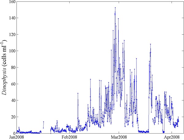
In mid-February, Campbell began to notice that the Imaging FlowCytobot was detecting rising levels of another toxic algae, Dinophysis cf. ovum, which causes diarrhetic shellfish poisoning in humans. Symptoms include nausea, cramping, vomiting, and diarrhea. Cooking the shellfish does not destroy the toxin.
“We had never before observed a bloom of Dinophysis cf. ovum at such levels in the Gulf of Mexico,” Campbell said.
In early March, Texas health officials closed local bays to shellfish harvesting and recalled Texas oysters, clams, and mussels. The Fulton Oysterfest, which attracts thousands of visitors, went on as scheduled March 6 to 8—by bringing in shellfish from non-tainted waters. No shellfish-related human illness was reported.
“We were looking for Karenia brevis and weren’t expecting to find a bloom of something else,” Sosik said. “No one necessarily would have looked for this algae until after people had gotten sick.”
“This is exactly what an early warning system should be,” Campbell said. “It should detect a bloom before people get sick. So often, we don’t figure out that there is a bloom until people are ill, which is too late. The Imaging FlowCytobot has proven itself effective for providing an early warning.”
Olson said it was “very satisfying to find that a technology we developed as a research tool can be so effective for protecting human health.” He and Sosik were recently awarded funding from the National Oceanographic Partnership Program to work with a commercial partner to design and build a version of the Imaging FlowCytobot for commercial use.
“This will ultimately enable more widespread use of this technology by scientists and resource managers,” Sosik said.
Funding for Campbell’s monitoring program and construction of the instrument was provided by the National Oceanic and Atmospheric Administration’s Cooperative Institute for Coastal and Estuarine Environmental Technology (CICEET).
Funding for instrument development and earlier prototypes of the FlowCytobot and the Imaging Flow Cytobot was provided by WHOI—through its Ocean Life Institute, Coastal Ocean Institute, Bigelow Chair, and Access to the Sea Fund—and by the National Science Foundation. The Gordon and Betty Moore Foundation has funded Olson’s and Sosik’s continuing work on automated submersible flow cytometry.
Slideshow
Slideshow
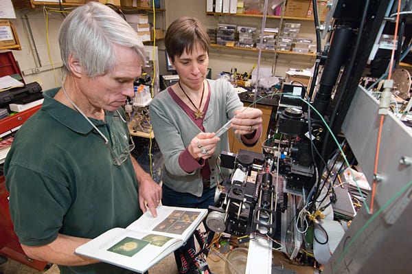
- Rob Olson and Heidi Sosik, biologists at Woods Hole Oceaonographic Institution, developed a device called the Imaging FlowCytobot, an automated underwater microscope that reveals plant and animal life in the ocean. (Photo by Tom Kleindinst, Woods Hole Oceanographic Institution)
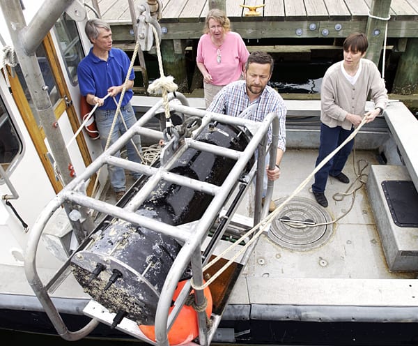
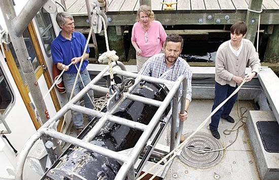
- Researchers lower the FlowCytobot, a foreruner of the Imaging FlowCytobot, onto the WHOI research vessel Mytilus. (Photo by Tom Kleindinst, Woods Hole Oceanographic Institution)
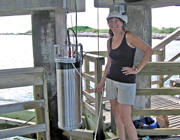
- In the fall of 2007, Sosik and Olson collaborated with biological oceanographer Lisa Campbell at Texas A&M University to deploy the Imaging FlowCytobot in the Gulf of Mexico to look for seasonal blooms of the toxic algae Karenia brevis. In mid-February, Campbell began to notice rising levels of an unexpected toxic algae, Dinophysis acuminata. (Courtesy of Lisa Campbell, Department of Oceanography, Texas A&M University )
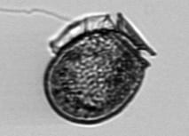
- The Imaging FlowCytobot captured this image of the toxic alga Dinophysis cf. ovum during the bloom in March 2008. (Heidi Sosik and Rob Olson, WHOI, and Lisa Campbell, Texas A&M)
- Data from the Imaging FlowCytobot shows the increase in abundance of Dinophysis cf. ovum in March 2008. (Heidi Sosik and Rob Olson, Woods Hole Oceanographic Institution)
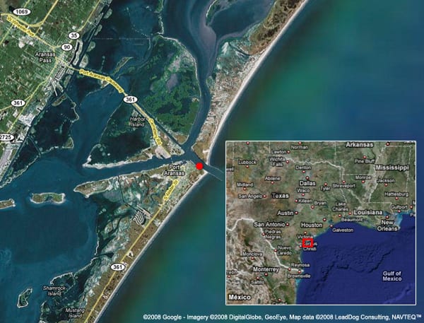
- The Imaging FlowCytobot was deployed off Port Aransas, nearCorpus Christi, Texas.
Related Articles
- Busting myths about HABs
- Are warming Alaskan Arctic waters a new toxic algal hotspot?
- Sargassum serendipity
- A dragnet for toxic algae?
- The Recipe for a Harmful Algal Bloom
- As Bay Warms, Harmful Algae Bloom
- Not Just Another Lovely Summer Day on the Water
- Setting a Watchman for Harmful Algal Blooms
- Dropping a Laboratory into the Sea
Featured Researchers
See Also
- Building an Automated Underwater Microscope An interview with WHOI biologist Heidi Sosik from Oceanus magazine.
- Heidi Sosik's Lab Optical Oceanography and Phytoplankton Ecology
- Rob Olson
- Lisa Campbell
- Revealing the Ocean's Invisible Abundance from Oceanus magazine
