Nathan Mollica (left), a graduate student in the MIT-WHOI Joint Program, and WHOI scientist Weifu Guo examine a sample cored from the skeleton of a coral. They put the cores in a 3-D Computerized Tomography (CT) scanner to image the coral skeletons. The CT images reveal annual growth bands, much like rings on a tree. The images helped the scientists identify a detailed mechanism showing how ocean acidification affects coral skeleton growth. It gives scientists a way to predict more precisely where corals will be more vulnerable. Mollica, Guo, and WHOI scientist Anne Cohen co-authored a study published in the Proceedings of the National Academy of Sciences. (Photo courtesy of Anne Cohen Lab, Woods Hole Oceanographic Institution)
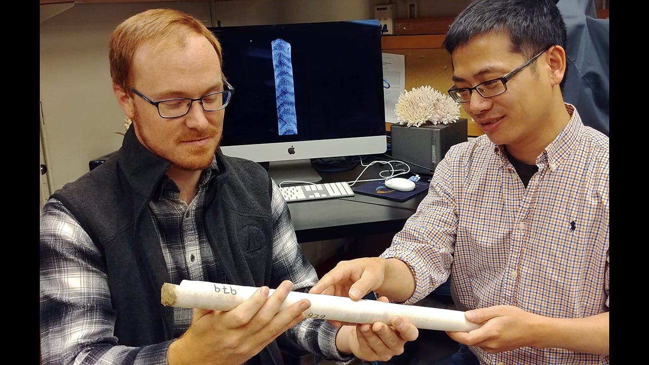
Image and Visual Licensing
WHOI copyright digital assets (stills and video) on this website can be licensed for non-commercial use upon request and approval. Please submit your request via our Media Request Form.
For assistance or accessibility accommodations, call (508) 289-2647.