 |
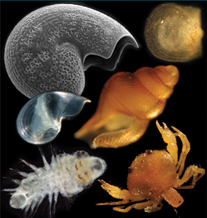
Photographic Identification Guide to Larvae at Hydrothermal Vents in the Eastern Pacific
|
| Introduction | Methods | Using This Guide |
Introduction
For animals living on the seafloor, a planktonic larval stage is a critical phase of the life cycle. Larval dispersal provides ecological and genetic connections among communities in patchy habitats such as hydrothermal vents. Temporal variation in larval supply to benthic communities can lead to fluctuations in the size and genetic composition of adult populations. On long time scales, barriers to dispersal can lead to speciation and are thought to be fundamental factors in generating biogeographic patterns and regional biodiversity. Despite the importance of the larval phase, very little is known about larval dispersal in the deep sea, even at hydrothermal vents where the habitat is patchy and transient, and larval exchange critical to the survival of endemic species.
General difficulties of larval identification for deep-sea studies include the scarcity of larvae in plankton samples, the fact that the adults may be unknown, and the difficulty of matching larval morphotypes to adult forms. However, some hydrothermal vent habitats have well-characterized benthic communities with relatively low species diversity and relatively high biomass and fecundity, resulting in large numbers of larvae in the plankton compared to typical deep-sea habitats. In addition, a large portion of hydrothermal vent communities can be comprised of gastropods, which can, in many cases, be identified by protoconch morphology. For example, gastropod larvae collected near hydrothermal vents in the eastern Pacific have been identified morphologically under light and electron microscopy (e.g. Mullineaux et al., 1996).
Since the discovery of hydrothermal vents thirty years ago, researchers have been collecting larvae in studies to explain the colonization of these oases in the deep (e.g. Lutz et al. 1984, Turner et al. 1985, Kim and Mullineaux 1998). Recent emphasis has been placed on time-series collections of larvae in multi-disciplinary studies of larval dispersal and supply to vent communities, such as the LADDER project at the East Pacific Rise. The purpose of this photographic identification guide is to serve researchers studying hydrothermal vent larvae in previously collected and future samples. The photographs may also be useful to those studying newly settled colonists.
Methods
Collection and preservation of larvae
For this first edition of the identification guide, larvae were collected near hydrothermal vents at the East Pacific Rise (EPR) 9° N site. Specimens were obtained over a 15-yr period, beginning with collection by nets and pumps with small-volume samples from 1991 – 1995 (Kim and Mullineaux, 1998), pumps with large-volume samples from 1998 – 2007 (Mullineaux et al., 2005; Beaulieu et al., 2009), and time-series sediment traps from 2004 – 2007 (Adams, 2007). We note that net tows, plankton pumps, and sediment traps do not sample larvae in equal proportions – some are better collected by one method or another and a combination of methods is likely to give a more complete description of the larval species composition of a particular site (Beaulieu et al., 2009).
For our recent studies at the EPR, large-volume pumps were used to collect discrete plankton samples over 1-day periods (McLane Large Volume Water Transfer System WTS-LV50; McLane Research Laboratories, Inc., Falmouth, MA, USA). We pumped at 30 L min-1 (500 cm3 s-1) over a filter comprised of 63µm Nitex mesh, yielding ~40 m3 pumped per day. For time-series sampling we used a conical, time-series sediment trap with sampling aperture 0.5 m2 and 21 cups (McLane PARFLUX Mark 78H-21 Sediment Trap; McLane Research Laboratories, Inc., Falmouth, MA, USA). Prior to deployment, we filled the cups with a solution of 20% dimethylsulfoxide (DMSO) in ultrapure water saturated with NaCl. We chose this preservative to allow for molecular genetic analyses of the collected specimens (e.g. Comtet et al., 2000). The pumps and sediment traps were deployed on autonomous subsurface moorings, with the samples collected between 2 and 175 m above bottom (mab) depending on each mooring configuration. Moorings were positioned within or near (< 2 km off-axis) the axial summit trough.
For the large-volume pump samples, after recovery on deck the filter holder was removed into a 20-L bucket with chilled, filtered seawater. All subsequent handling of the sample occurred in a cold room (4º C). Samples were carefully rinsed from the filter using a squirt bottle with chilled, filtered seawater. Many of the collected specimens were alive upon retrieval of the pump. We briefly examined the samples live under a dissecting microscope prior to collecting onto a 63µm sieve, rinsing with fresh water, then preserving in 95% ethanol for examination at our laboratory. For the sediment trap samples, after recovery of the mooring we photographed the cups and stored them at 4º C prior to shipment to our laboratory for examination.
Sorting and photographing larvae
For sorting at our laboratory within a few months after each cruise, samples were poured over nested 300µm and 63µm sieves, and each fraction was rinsed with fresh water into a petri dish. We sorted larvae under a dissecting microscope at 25X, with identification generally at 50X; some specimens required examination under a compound microscope at 100X. Individual larvae were manipulated with a fine paintbrush or short length (~5 mm) of human hair glued to the end of a wooden stick. Individuals were transferred with a pipette set to ~10µL. For examining under the compound microscope, individuals were transferred to a welled slide filled with fresh water. We moved the cover slip gently side-to-side to roll the larva into an appropriate position for measuring and photographing. Larvae sorted from both sediment trap and pump samples were saved in 95% ethanol and stored in Lauren Mullineaux’s laboratory at Woods Hole Oceanographic Institution. We do not recommend transfer from DMSO solution to ethanol for future studies because it apparently caused tissue degradation for polychaete larvae.
Some gastropod larvae were dried and imaged using scanning electron microscopy (SEM). These specimens were placed on ½” diameter circular cover slips which had been previously coated with a thin layer of white Elmer’s glue, which was allowed to dry. The small amount of ethanol clinging to specimens dissolved the glue enough to stick them in place. The cover slip with the specimen was then attached to a SEM stub and sputter-coated for 1 min using Samsputter. These were examined using the JEOL 840 scanning electron microscope at the Marine Biological Laboratory (MBL).
Species identifications were made using a variety of sources. For gastropod protoconchs we relied heavily on the literature, which contains many detailed SEM photographs of protoconchs for most of the EPR 9° N species. For species for which the protoconch is unknown, we occasionally would image the protoconch of an identified juvenile for comparison (e.g. Gorgoleptis spiralis, Adams et. al., 2010). For the identification of Bythograea sp. zoea, we are indebted to Ana Dittel (University of Delaware), who examined one of our specimens. Our mussel larvae were all at a stage near to settling and could be directly compared to the shells of settled juveniles. For polychaete larvae, we were forced to use comparisons with newly settled juveniles or similarity to larvae of other, related species; thus, most of these could not be identified to species. Assignment of deep-sea polychaete larvae to species awaits development of molecular genetic probes (e.g. Pradillon et al., 2007).
Most of the photographs in this guide were taken through a Zeiss Axiostar compound microscope under brightfield, usually at 100X and occasionally only at 50X for larger specimens. For most of the photographs we used a Nikon D100 SLR digital camera with resolution 3008 x 2000 pixels and twelve bit dynamic range. We used an AF Zoom-NIKKOR 28-200mm lens and obtained the best images with F-stop 8, at shutter speed ~1/125 sec. Some photographs were taken with the same camera and a Zeiss Stemi 2000-C dissecting microscope. A few photographs were taken using a Zeiss Discovery.V12 Axiovision system dissecting microscope and Zeiss Axiocam MRc5 camera with Axiovision software at the MBL. The original images were cropped, scaled to approximately 400 x 400 pixels (~520 x 400 for SEMs) for the higher definition pictures, and adjusted using Adobe Photoshop software to enhance details.
For those who would like more information about methods of preserving, handling and storing small gastropods, we highly recommend Geiger et al., 2007.
Using this guide
This guide is intended to serve as a reference for the morphological identification of larvae collected near hydrothermal vents. In this first edition, the species are restricted to those found at EPR 9º N. However, in future editions of this guide, we would like to include hydrothermal vent larvae from other regions, and we encourage contributions to our website. We organized the guide into two general sections: 1) gastropod larvae, and 2) larvae of other invertebrate taxa. To use this guide to identify larvae, we recommend using both a dissecting and compound microscope, each with a calibrated micrometer in the eyepiece.
Standard Description Format
Top line: Species name (or morphotype), family, and original reference for description of the species.
Photo panels: Light microscopy, followed by SEM for gastropod protoconchs and, often, dark field for polychaete larvae.
Additional references with photographs are listed below the photo panels.
Size: Provided for calibrated dissecting and compound microscopes.
Morphology: Gastropod protoconchs and polychaete larvae are described using standard terminology (see Terminology page). If the morphotype is not identified to species, this section lists references to similar-looking species.
Frequency: For frequency designations, we used larval abundance data
at EPR 9º N for on-axis, near-bottom pump samples from 1998-2000 (4 cruises, 4 locations, 12 samples; subset of data in Mullineaux et al., 2005) and 2004 (1 cruise, 1 location, 5 samples;
Beaulieu et al., 2009) and one on-axis, near-bottom deployment of a time-series sediment trap from Nov. 2004 - Apr. 2005 (Adams, 2007).
We used four categories to describe the frequency of each species (or morphotype):
Common: Present in majority of samples, at relatively high abundance overall (i.e. >5%)
Frequent: Present in majority of samples, at relatively low abundance overall (i.e. <5%)
Occasional: Present in < ½ samples, at variable (usually low) relative abundance per sample
Rare: Present in very few samples, with only a single individual per sample.
Table for: “Can be confused with": This section provides a list of similar-looking species (or morphotypes), with comparisons of morphological features and thumbnail images to click open in a pop-up window.