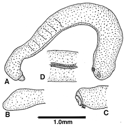
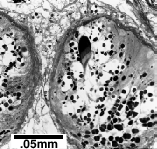
Figure
left: A entire holotype, note protruded genital cone. B dorsal posterior
view. C fold around mouth, long cilia from pedal pit. D pedal groove spicules,
anterior to right. Figure right: holotype
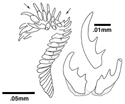
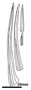
Copulatory
spicules (figure left) hollow, fluted, curved, in paired bundles numbering
up to 15 spicules per bundle varying in length from 15 to 200 µm,
the shorter with the same morphology as the distal ends of the longer ones
(figure left).
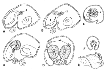
A-F,
semischematic drawings of reproductive system from histologic sections,
anterior (A) to posterior (F). G, posteroventral view of individual with
cuticle turned back from shallow mantle cavity showing free proximal ends
of copulatory spicule sacs. 1 seminal receptacle, 2 lower gametoduct, 3
copulatory spicule sac, 4 upper gametoduct, 5 exterior of genital cone.
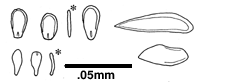
Most
epidermal spicules rimmed, nearly oval, somewhat asymmetrical, narrowest
at flattened base, with medial bump, to 25 µm long by 14 µm
wide; curved paddle-shaped spicules few, 20 by 9 µm; spicules beside
pedal groove (at right) 38 to 58 µm long to 16 µm wide, overlapped
in flat, paired ventral longitudinal rows; all spicules <2 µm
thick.
Appearance
mossy, height <0.5 mm, body narrowing posteriorly before broadening
at posterior end, width greatest ventrally (A-C). Cuticle <2 µm
thick, epidermis to 11 µm thick; anterodorsal midgut coecum paired,
large; with a pedal commissure sac anteriorly (Scheltema, 1981).
Radula
0.2 mm long with 20 to 30 rows, those near proximal end opening into pharynx,
distal half held vertically in single anteroventral radular pocket, teeth
30 to 35 µm long with 2 median denticles.
Reproductive
system. -- The paired upper gametoducts are short and enter the lower
gametoducts dorsolaterally presumably shortly after the dorsomedial union
of the lower gametoducts with the pair of large, ventral, lobate seminal
receptacles (B, C). The upper gametoducts were clearly distinguished in
3 sectioned individuals (D), but neither their origins from the pericardial
cavity nor their union with the lower gametoducts were discerned. The paired
lower gametoducts are surrounded by circular muscles, which become more
pronounced after fusion of the two gametoducts to form a cone (G) similar
to that in the genera Genitoconia Salvini-Plawen (1967) and the
lepidomeniid Nierstrassia Heath (1918, pl. 6 fig. 11). The mantle
cavity runs as a shallow space beneath the lower gametoducts, genital cone,
and copulatory spicule sac (D). The proximal ends of the copulatory spicule
sacs lie free in the mantle cavity (A-C, G); distally they lie within the
body cavity (D, E). The relationship of the openings of the copulatory
spicule sacs and the genital cone into the mantle cavity were not clear
from the sections. The copulatory spicules are apparently deciduous, with
new spicules forming in a rosette of cells within the copulatory spicule
sacs (see Scheltema et al., 1994, fig. 23D). Spermatophores, not reported
in any other species of Aplacophora, occur in the lower gametoducts and
genital cone (figure above).
In G. pellucida, the union of the lower gametoduct and seminal
receptacle was figured but interpreted as the union of upper and lower
gametoducts ( Odhner, 1921, fig. 85); the upper gametoducts, which are
difficult to see, were not figured.
Remarks.
--
Gymnomenia virgulata is placed in the genus
Gymnomenia
on the basis of body shape, striped appearance due to the midgut sacculations,
mouthfold of everted pharynx, and structure of the gametoducts. It is distinguished
from species belonging to
Wirenia Odhner (=
Aesthoherpia Salvini-Plawen,
1985; see Salvini-Plawen, 1997) by spicule morphology and from
Genitoconia
Salvini-Plawen by the separation of mouth and vestibule and by the presence
of midgut sacculations.
G. virgulata differs from
G. pellucida
in height, which is greater and even throughout the body in
G. pellucida,
in number of copulatory spicules, and in presence of a large, paired midgut
coecum, which is small and single in
G. pellucida.
A pedal commissure sac with its contained bodies found in
Gymnomenia
also occurs in
Genitoconia and
Wirenia. Its fine structure
has been described in
Wirenia (Haszprunar, 1986 [as
Aesthoherpia
]; Scheltema et al., 1994).
From Ophelia 51 (1): 1-28 (1999).
Gymnomenia
virgulata Scheltema, 1999
Gymnomenia sp., Scheltema, 1981, figs. 2m, n, 4a-g; Scheltema
et al. 1994, figs. 13c, 23b, d, 24a.
Type locality. -- Off Walvis Bay, Namibia, 23°00'S, 12°58'E,
619-622 m (RV ATLANTIS II Cruise 42, Stn. 188, 16.v.1968).
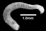
Holotype. -- USNM 880314 (alcohol specimen, spicule slide). 4.1
mm long, height 0.4 and 0.3 mm anteriorly and posteriorly, respectively
. Paratype 1. -- USNM 880315 (dissected alcohol specimen, spicule slide
including copulatory spicules). Paratype 2. -- USNM 880316 (alcohol specimen).
Paratype 3. -- USNM 880317 (radula slide only). All from type locality.
Etymology. -- gymno = bare; virgulata = stripes