Acanthomenia
arcuata Scheltema, 1999
Acanthomenia sp., Scheltema et al. 1994, fig. 5f.
Type locality. -- West European Basin, 55°07.7'N, 12°52.6'W,
2,897 m (INCAL [CENTOB] DS-09, 20.vii.1976).
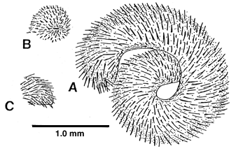
Holotype. -- MNHN (alcohol specimen, spicule slide). Length
4.7 mm, midbody height 0.6 mm. Paratype 1. -- MNHN (radula and spicule
slides). Type locality .Etymology. -- acantho- = thorn; arcuata
= bent like a bow.
A
small, very spiculose species to 6.4 mm long, height even throughout, to
0.7 mm; anterior end rounded, contracted mouth opening a short, horizontal
slit; posterior end pointed ventrally in lateral view. Cuticle 11 µm
thick, epidermis 22 µm thick.
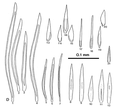
Hollow
epidermal spicules curved, recurved at base and usually at thick, solid
tip, long spicules to 340 µm long by 14 µm wide, length of
solid tip to 35 µm, length of solid base to 14 µm, walls of
hollow section 2 to 3 µm thick (spicules 1-4); also many short, narrow
hollow spicules (spicules 5, 6). Solid spicules of two types: one type
widest and thickest (to 6 µm) at midpoint, distal end gradually tapered
to point, to 124 µm long by 14 µm wide (spicules 8-12); second
type often asymmetrical, shorter, to 100 µm, very thin (2 µm),
pointed, and rimmed around base, some with narrow, drawn-out tip (spicules
13-18). Pedal groove lined on either side by thin, basally rimmed solid
spicules with a basal stem (spicules 19, 20); lateral to these are slender,
curved hollow spicules 150 µm long by 7 µm wide (spicule 7).
A medial, platelike structure of numerous small (52 by 9 µm), thin
(1 µm), pointed spicules with rimmed edges and straight bases aligned,
present at mantle cavity opening (spicule 21).
Radula
small, monostichous, rows few, teeth about 70 µm long by 50 µm
wide (Figs 3A, B), most recent tooth with evidence that teeth are formed
by fusion of a pair of teeth onto a single base, each pair in turn with
two fused denticles; older teeth without indication of fusion of paired
teeth to base.
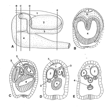
Reproductive
system. -- Acanthomenia gaussiana Thiele was originally described
from a single juvenile. In mature A. arcuata (A and sections B-E) the gonopericardial
duct is single as is the lower gametoduct, which, however, has a pair of
anterodorsal lobes (B). Eggs are produced as usual along the medial septum
between paired gonads; spermiogenesis is restricted to the posterior region.
The paired upper gametoducts join the lower gametoduct dorsally on the
dorsal lobes (C); shortly posterior to this union the dorsal lobes pinch
off and the upper gametoducts bend sharply ventrally, receiving the openings
of the large, paired, tubular, posteriorly extended seminal receptacles
(D). The lower gametoduct continues to narrow posteriorly (E), emptying
into the mantle cavity as a tube. The small pericardial cavity bends abruptly
ventrally just beyond the posterior ends of the seminal receptacles, merging
with the origins of the paired upper gametoducts. No copulatory spicules
or copulatory spicule sacs are present; however, the plate of tiny spicules
lying medially at the opening of the mantle cavity may be implicated in
copulation.
Remarks.
--
Acanthomenia arcuata differs from
A. gaussiana Thiele
in form of the epidermal spicules (Thiele, 1913, text-fig. 2) and of the
radula, known only from histologic sections (Salvini-Plawen, 1978, fig.
150). Individuals of
A. arcuata are so spiculose and curled up that
the forms of the mouth, pedal pit, and mantle cavity opening are difficult
to distinguish. To prepare radulae for examination it was necessary to
remove spicules with acid before the buccal mass could be extracted.
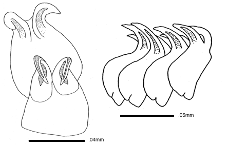
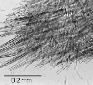
Figure right: Although skeletal spicules are not present, short,
solid adpressed spicules lie at an angle to and beneath the erect long,
hollow spicules. They thus behave structurally like skeletal spicules,
which by definition lie layered within the cuticle.
From Ophelia 51 (1): 1-28 (1999).
A,
reconstruction from histologic sections, anterior to left. B-E, cross-sections
indicated in A, semischematic. 1 pericardial cavity, 2 seminal receptacle,
3 upper gametoduct, 4 lower gametoduct, 5 midgut, 6 intestine, 7 heart.